Horses are very dependant on vision both as instinctive prey animals and as performance horses in which ever field of competition they are involved. While many ocular diseases in the horse have the potential to heal if treated they can just as easily deteriorate to cause irreversible loss of vision. Therefore all conditions of the eye require veterinary examination to ensure the best outcome.
Common eye problems
Eye disease in the horse represents a significant component of equine veterinary practice. The equine eye is prone to problems due to its anatomy, with a large orbit protruding from the side of the head, and decreased immunity to disease. Equine ophthalmic disease, if inadequately treated can lead to loss of vision.
The equine eye is made up of the upper and lower eyelids that are covered with skin, the third eyelid or “nictitating membrane”, the lacrimal or tear system, the cornea and conjunctiva, the anterior chamber, the iris and pupil, the lens and ciliary body, the vitreous and the retina and optic nerve. Problems can occur in any part of the eye resulting in disease and there is the potential for severe damage which may result in the loss of vision.
Clinical Signs of Eye Disease
Eye disease in the horse may manifest in many ways. A horse showing any or a combination of the following signs should be examined by a veterinarian as a matter of urgency:
- Pain, which leads to partial of complete closure of the eyelids
- Excessive tear production
- Swelling of eyelid/s
- Redness
- Rubbing at eye
- Change in corneal appearance. A normal healthy cornea is clear and an unhealthy one may become blue/cloudy because of oedema or cellular infiltrate.
- Differences in pupil size between eyes
- Flecks of material within the fluid in the eye.
Common Conditions
In equine practice, corneal disease or injury (damage to the surface of the eye) is probably the most frequent reason eyes are examined. This is largely a consequence of the degree of exposure of the equine cornea to potentially harmful agents. While the eyelids and lacrimal system work to protect the eye, it is the cornea that has the most important barrier function, protecting the deeper structures of the eye from micro-organisms.
The normal horse cornea is very thin, measuring a total of less than 2mm deep and consists of five layers:
- tear film
- corneal epithelium
- corneal stroma
- descemet’s membrane
- endothelium
Positive stain of a deep ulcer within a shallow ulcer (ulcerated area shown in green dye)
Fungal Stromal Abscess (cream dot in eye circled in red)
A corneal bulla
Corneal Ulcers
A corneal ulcer occurs when there is loss of corneal epithelium. There are many common causes of corneal ulceration including direct trauma, foreign bodies, eyelid dysfunction, eyelid tumours, and abnormal or extra growth of eyelashes that may scratch the cornea. The most common cause however is a scratch to the surface of the eye. Horses can scratch their eyes easily when grazing in tall grass or amongst shrubs, or if dirt is thrown in their face by another horse as seen with race horses during competition. This results in a superficial ulceration of the cornea. These simple, uncomplicated corneal ulcerations usually heal in less than seven days, however many of these ulcerations are complicated by infection. Many bacteria and fungi live normally on the surface of a horses’ eye as a consequence of the grazing behaviours and the environment they live in. These bacteria cause no harm to the eye unless they infect one of these simple uncomplicated ulcers. The consequence of this infection is an otherwise simple scratch to the eye becoming a potentially vision threatening condition. Infection with bacteria or fungi results in the ulcerated area of the cornea becoming deeper. The real risk with severe infections occurs when the ulcer extends through the full depth of the cornea, rupturing the eye.
All corneal ulcers require urgent veterinary attention and intervention. The aim of treating an uncomplicated simple ulcer is to prevent infection taking hold while the cornea heals. Conversely the aim of treating a complicated ulcer is to aggressively attack the infecting bacteria and fungi. This may involve treatment every two hours around the clock for severe cases.
Melting Ulcers
If infection and sever inflammation are not controlled, a deep complicated ulcer can quickly become a “melting ulcer”. The term “melting ulcer” is used to describe an uncontrolled infection or inflammatory response in the stroma leading to liquefaction of the cornea. Melting ulcers are not uncommon in horses and constitute a genuine ocular emergency because of how quickly the cornea may perforate.
Herpes Keratitis
The only recognised infectious primary cause of corneal ulcers in horses is herpes virus. Herpes Virus infection can lead to multiple pinprick ulcers developing in the cornea. These are treated with topical antiviral medication.
Stromal Abscess
A stromal abscess occurs when a small pin prick ulcer becomes infected with bacteria and/or fungi. The ulcer heals quickly, trapping the infection within the deep layers of the cornea, the stroma. These are different to infected ulcers as the epithelium of the cornea is intact. The initial abscess is so small as to not be visible with the naked eye. As the infection progresses, the abscess increases in size appearing like small spots of white/yellow in the cornea. The cornea immediately surrounding the abscess becomes cloudy and the eye is extremely painful. Treatment of stromal abscesses is challenging as many medications struggle to penetrate into the abscess. Medical treatment of stromal abscesses may take up to two to three months and a treatment tube should be inserted to ensure easy medication. Occasionally surgical intervention is indicated.
Bullous Keratopathy
Bullous keratopathy is a non-specific diagnosis that describes the formation of severe corneal oedema that gives the appearance of the cornea hanging over the lower eye lid margin. This severe oedema results either from an extensive shallow ulcer that allows fluid to enter the corneal stroma from the tear film or from blunt trauma damaging the endothelium allowing water to penetrate from the fluid inside the eye. It is managed as an intensive medical condition.
Uveitis
Uveitis is defined as inflammation of the internal structures of the eye. Uveitis is extremely painful in the horse and commonly occurs secondary to any ocular injury or disease. It can however occur as a primary entity.
Uveitis is a very important diagnosis in equine veterinary practice as it may lead to irreversible blindness if untreated. It is the most common cause of blindness in horses and Appaloosa are genetically predisposed to the disease. Treatment of uveitis includes identifying an underlying disease, if one is present eg. corneal ulceration. If there is no obvious primary disease, then symptomatic treatment of uveitis is necessary to relieve pain and reduce the chance of permanent damage to the eye. It may be that the uveitis is secondary to a systemic illness that may need to be worked up by a veterinarian.
If vision is lost or the prognosis for a cure hopeless, the major goals of treatment include eliminating pain, minimising owner expense and effort. Often the best way to achieve this is by surgically removing the eye. Enucleation has very few patient side effects and leaves the patient comfortable once surgical pain has subsided.
Squamous Cell Carcinoma
Squamous cell carcinoma is a common tumour in horses, especially in less pigmented horse breeds eg. Paints, Appaloosas. Lesions may be found on the eyelids, including third eyelid. Early in the disease process signs of low grade inflammation may be noticed eg. slight mucous discharge, slight reddening. As the carcinoma grows it may appear more vegetative-like and may eventually ulcerate. Squamous cell carcinoma lesions on the third eyelid can be treated by surgical excision and/or cryotherapy (freezing). This can be performed in a standing sedated horse or under general anaesthesia. If the lesion is quite deep, entire removal of the third eyelid may be warranted.
Habronema
Another important disease of the eye is a habronema infection. This occurs when flies lay eggs onto conjunctiva which then develop into microscopic maggots. Initially the infected eye may appear like conjunctivitis, and veterinary consultation will diagnose the infection. Treatment includes physical removal of the infection followed by application of specific mediation. Regular worming with Ivermectin helps prevent this disease.
Conjunctivitis
Conjunctivitis is a common term that most people use to describe a red and irritated eye. It actually means inflammation of the conjunctiva and can be caused by bacterial, viral or parasitic infection, irritation from flies or even a dusty environment. An eye with conjunctivitis should not be painful. While it may appear that conjunctivitis is common and not en emergency, it is often a symptom of another underlying eye disease and so a veterinarian should be consulted.
Eyelid Lacerations
Horses commonly injure their eyelids. It is essential that all efforts are made to repair damage to the eyelid margin as failure to do so can result in corneal irritation and pain as well as a chronic weepy eye.
Eyelid Tumours
Many tumours may arise on or near eyelids. Any of these tumours that impinge on the cornea in any way will result in chronic irritation and ulceration. Eye lid tumours are treated by surgical removal and or chemotherapy. It is essential that early intervention be considered as removal of a larger tumour can result in eyelid defects.
Treatment
Treatment of eye disease may be categorised as medical or surgical. Treatment depends on the specific condition, however generally involves:
- Resolution of the primary cause of disease eg. foreign body removal.
- Broad spectrum antibiotic/ antifungal directly to affected eye.
- Pain management and anti-inflammatories eg. systemic NSAID’s; any insult to the corneal epithelium is extremely painful for the horse.
- Topical atropine; Even minor damage to the cornea can cause a reflex uveitis, therefore dilating the pupil is an important part of treatment.
Surgery may be indicated with deeper corneal ulcers to help protect the cornea, promote healing and lessen the risk of perforation. Surgical options include: conjunctival grafting, third eyelid flap and temporary tarsorraphy.
Conjunctival grafting is performed under general anaesthesia. The conjunctiva is surgically transplanted onto the defective cornea with a small flap or the conjunctiva can be loosened and pulled to cover the entire cornea. These grafts provide a direct blood supply to the cornea and hence increase its healing ability. These grafts also provide structural support to areas of extremely thin cornea. Once the cornea has healed, the graft may be surgically trimmed to reduce the area of scarring.
Performing a temporary tarsorraphy is a common procedure where the eyelids are sutured together temporarily to provide protection for the diseased eye. Although it may sound simple, this technique helps the healing process dramatically. If the eyelids are sutured together, it is more difficult for medication to be applied directly to the eye, therefore a treatment tube may also be necessary.
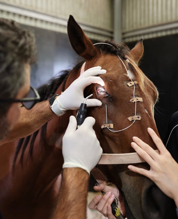
Treatment tubes are of considerable value when frequent or long term topical treatment of the eye is required. They can be inserted in the sedated conscious horse using local anaesthesia, or under general anaesthesia following surgery. A treatment tube consists of a plastic footplate which surrounds a small central hole at the end of a plastic delivery tube. The footplate sits under the eyelid and allows direct application of medication to the eye. The main hurdle to effective medication of the horses’ eye is the horse itself. Eyes requiring such intensive treatment are painful and so it can be very difficult if not impossible to effectively and consistently apply the required medication. Treatment tubes make it possible to medicate the painful eye with minimal contact.
By Dr Caitlin McGuckin BVSc (Hons)
Medication tube inserted into horses eye with tarsorraphy (eyelids sewn shut to help protect the eye)
